
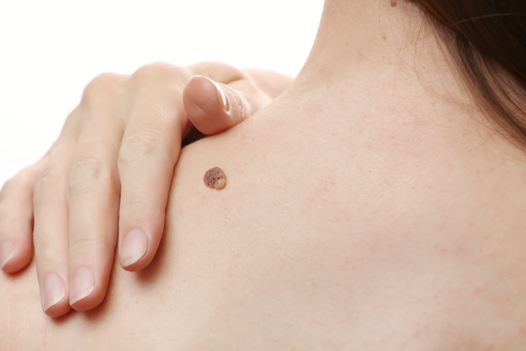
How Mohs removes cancerous cells layer by layer while preserving as much healthy tissue as possible — the gold standard for many skin cancers.
What Makes Mohs Different
Mohs micrographic surgery removes skin cancer one thin layer at a time. Each layer is examined under the microscope the same day. The surgeon continues only where cancer cells remain — sparing healthy tissue and confirming clear margins before you leave.
When Mohs Is Recommended
Mohs is often chosen for cancers on the face, ears, nose, lips, hands, and other areas where preserving healthy skin matters most. It is also used for recurrent tumors, aggressive subtypes, and cancers with poorly defined borders.
What To Expect On Procedure Day
Most Mohs visits take a half day. You arrive, the visible tumor is removed, tissue is mapped and examined, and additional layers are taken only if needed. Bring a book, wear comfortable clothes, and plan to wait between stages while the lab works.
Reconstruction And Healing
Once margins are clear, the site is repaired with care for both function and appearance — sometimes the same day, sometimes with a staged plan. Dr. Wall is fellowship-trained in Mohs and reconstructive surgery, so precision does not stop at tumor removal.
Cure Rates You Can Trust
For many common skin cancers, Mohs offers among the highest cure rates of any treatment — often cited above 99% for primary basal cell carcinoma — while keeping scars as small as the cancer allows.
Before You Go
No. Mohs is most often used for basal cell and squamous cell carcinomas. Melanoma care follows different protocols; your dermatologist will guide the right approach.
Many sites are closed with sutures or a reconstructive technique suited to the size and location. Some small sites may heal by secondary intention.
Most patients resume light activity within a few days. Full healing depends on the site and repair — we provide clear aftercare for every step.
Reading is a start. A visit is an answer.
Schedule An Appointment →