
Mohs Skin Cancer Surgery
Remove the cancer. Preserve healthy skin. Reconstruct with intention.
Skin Cancer Removal, Under The Microscope.
Unlike a standard excision, Mohs surgery combines surgical removal and microscopic examination during the same visit.
Mohs surgery is a specialized technique for treating certain skin cancers. The tumor is removed along with a thin layer of surrounding tissue. That tissue is carefully mapped, processed, and examined under the microscope.
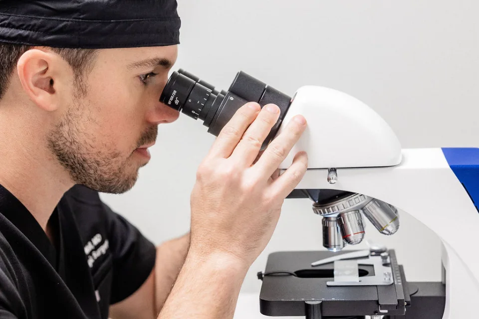
If cancer cells remain at a margin, the map tells the surgeon precisely where to remove additional tissue. The process is repeated only where cancer remains — until the margins are clear upon examination.
This ability to evaluate margins during surgery allows Mohs to achieve a high cure rate while preserving as much unaffected skin as possible — an important consideration for the nose, eyelids, ears, lips, hands, and other functionally or cosmetically important locations.
Remove the cancer. Preserve healthy skin. Reconstruct with intention.
Precise Removal. Maximum Preservation.
Mohs surgery isn’t necessary for every skin cancer. When it is indicated, its strength lies in combining margin control and tissue preservation.
Each layer is mapped and examined during surgery, so additional tissue is only removed specifically where cancer remains.
By taking additional tissue only where needed, Mohs helps preserve surrounding healthy skin.
Mohs offers a high cure rate for appropriately selected skin cancers, including certain tumors with a higher risk of recurrence.
Once the cancer is cleared, attention turns to the wound — its location, size, depth, surrounding anatomy, function, and the best approach to healing or reconstruction.
How Mohs Surgery Works.
Patients remain awake throughout — the treatment area is numbed with local anesthetic. Click through the four stages.
The Mohs surgeon removes a thin layer of skin from the visible cancer site. This tissue is immediately prepared for microscopic examination in our on-site lab.
The removed tissue is examined under a microscope to check for cancer cells at the edges — the “margins.” This critical step lets the surgeon see exactly where any remaining cancer cells are located.
If any cancer is seen at the margins, another very thin layer is removed only from the areas where cancer cells were found. This is repeated layer by layer, focusing only on areas with cancer, until all cancerous cells are gone — preserving as much healthy skin as possible.
Layer by layer. Only where needed.
Once all cancer is removed, Dr. Wall will often perform reconstruction right away to ensure the best cosmetic outcome — especially for areas like the face, where both appearance and function are incredibly important.
Ask about Mohs surgery close to home.
The Cancer Determines The Treatment.
Not every basal cell carcinoma or squamous cell carcinoma requires Mohs surgery. Your dermatologist considers the type of cancer, location, size, borders, prior treatment, recurrence risk, and other clinical features — Mohs may be particularly valuable when preserving healthy tissue or achieving detailed margin evaluation is important.
Face · nose · eyelids · ears · lips · hands · other anatomically important sites.
A tumor that has returned after previous treatment.
When it is difficult to determine where the tumor ends clinically.
Where removing unnecessary healthy tissue may meaningfully affect reconstruction, function, or appearance.
Skin cancers whose clinical or pathologic characteristics make Mohs an appropriate option.
Removal Is Only Part Of The Work.
Mohs surgery brings together several disciplines in one procedure: cutaneous oncology, surgery, microscopic interpretation, pathology, and reconstruction.
After dermatology residency, physicians pursuing accredited fellowship training in Micrographic Surgery & Dermatologic Oncology undergo an additional competitive fellowship lasting one to two years — with structured operative training and exposure to outcomes, recurrences, and complications.
Weston Wall, MD, FAAD, FACMS
Board-Certified Dermatologist · Fellowship-Trained, Board-Certified Mohs Surgeon
Dr. Wall performs the Mohs procedure and, when appropriate, the reconstruction that follows — removal and repair considered as parts of the same surgical plan.
Treatment begins with understanding the tumor’s pathology, location, and behavior.
The removed tissue is mapped and microscopically examined to identify where cancer remains.
Additional tissue is removed only from areas where residual cancer is identified.
After clearance, the defect is evaluated according to its anatomy, size, depth, tension, and surrounding tissue.
Cancer Clear. Now We Think About Healing.
Not every Mohs wound requires the same repair.
Some wounds may be allowed to heal naturally. Others can be closed in a straight line. More complex defects may benefit from movement of nearby skin using a flap or, in selected cases, a skin graft.
The appropriate approach depends on much more than the size of the wound — Dr. Wall considers location, depth, nearby structures, skin movement, tension, function, natural contours, and the eventual position of the scar.
The goal isn’t simply to close a hole. It is to choose a repair appropriate for the individual wound and surrounding anatomy.
Bring the wound edges together when appropriate.
Reposition nearby tissue while maintaining its blood supply.
Transfer skin from another location when appropriate.
Allow selected wounds to heal naturally without surgical closure.
Surgery Is One Day. Healing Takes Time.
Your care doesn’t end when the cancer is removed. A history of skin cancer increases the importance of continued skin surveillance and sun protection.
Asked & Answered.
Mohs is commonly used for selected basal cell carcinomas and squamous cell carcinomas and may be appropriate for certain other skin cancers. The diagnosis, location, and characteristics of the tumor determine whether Mohs is appropriate.
Yes. Mohs is performed in an outpatient setting using local anesthesia.
The amount of time varies. Each removed layer must be processed and examined, and some cancers require more than one stage. Patients should generally plan for the visit to take several hours.
Any surgery that cuts the skin creates a scar. Once the cancer is completely removed, the reconstruction is planned considering the location, anatomy, function, and eventual scar.
Dr. Wall performs reconstruction following Mohs when appropriate. The recommended repair depends on the individual defect and surrounding anatomy.
No. Different skin cancers and clinical circumstances call for different treatments. Mohs is recommended when its particular advantages suit the tumor.
Accredited Micrographic Surgery & Dermatologic Oncology fellowship training occurs after dermatology residency and provides additional structured training in Mohs surgery and related care.
No additional Mohs layers are required once the examined margins are clear. Attention then turns to wound management or reconstruction.
Continued dermatologic surveillance is an important part of care after a skin cancer diagnosis. Your dermatologist will recommend follow-up based on your individual history and risk.
From Diagnosis To Recovery.
Diagnosis
A biopsy identifies the type of skin cancer.
Treatment Plan
Your tumor’s pathology, location, and other characteristics determine whether Mohs is appropriate.
Mohs Surgery
The cancer is removed in stages with microscopic margin examination during the procedure.
Reconstruction
Once the examined margins are clear, the wound is evaluated and repaired when appropriate.
Follow-Up
Healing, scar maturation, and ongoing skin surveillance continue after surgery.
Skin Cancer Care, Close To Home.
A lesion of concern or a routine screening — from diagnosis through surgery, reconstruction, and follow-up.